Electron Microscopy - Principles
Electron microscopy is an imaging analysis method that can reach magnifications beyond optical microscopy. It can be systemised depending if the specimen is investigated using reflection of transmission of electrons and if the electron beam is scanning the specimen surface or not.
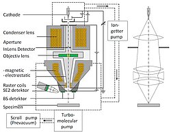
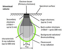
The most common Scanning Electron Microscopy uses electrons that are reflected from the specimen surface and slightly underneath the surface while scanning the specimen during an investigation. The maximum reachable resolution typically is between 1 nm and 10 nm. The electron beam can be generated using different methods, e.g. thermionic emission or by so-called Schottky emitters. Electrons then are accelerated in the electron tube using acceleration voltages up to some 10 kV, impact on the surface and penetrate the specimen to a depth of approximately 1 µm. Different interaction mechanisms of the electrons with the specimen materials generate backscatter electrons, secondary electrons, X-ray particles and others. Depending on the equipment stage, there are one or more detectors for each of those quantum particles, which are useful for different investigations based on their different principles. All those investigation have to be done in a vacuum chamber.
Special detectors allow analyses of specimen not only be acquiring images. One example is the Energy Dispersive X-ray Spectroscopy (EDX). Exposing the specimen surface with high-energy electrons material specific X-ray particles are generated. By evaluating the X-ray spectrum one can make conclusions about the existing kinds of atoms in the focus area of the electron beam.
Using the Electron Backscatter Diffraction (EBSD) it is possible to make the crystal structure of the specimen’s surface visible by so called diffraction pattern. Using software algorithms it is possible to analyse the crystal symmetry, the orientation, the location and boundary of crystal grains and much more.